Axillary and inguinal erythrasma - CMAJ
A 50-year-old man presented to the dermatology department with a 1-year history of itchy axillary and groin lesions. He had been treated with a topical antifungal preparation (cyclopyroxolamin), without improvement, by his family physician, who had suspected fungal intertrigo. On physical examination, we observed well-circumscribed, erythematous, brownish, scaly plaques affecting both armpits and the groin area bilaterally (Figure 1). To rule out superficial mycoses, we examined skin scrapings from the infected site under direct microscopy after potassium hydroxide preparation. We did not find any signs of fungal infection and did not isolate any dermatophytes in Sabouraud agar.
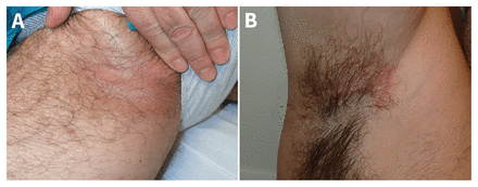
Photographs of a 50-year-old man showing erythematous, brownish scaly plaques affecting (A) groin and (B) armpit.
We suspected bacterial infection. Evaluated under Wood's lamp, the lesions exhibited coralred fluorescence (Figure 2). Gram staining confirmed erythrasma, showing clusters of bacilli in epithelial cells; Corynebacterium minutissimum was isolated in culture. We prescribed topical application of erythromycin gel twice a day, and the skin lesions resolved in 2 weeks.
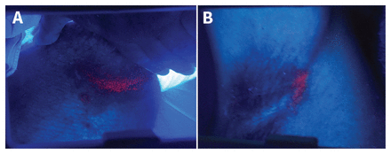
Photographs of (A) groin and (B) axillary lesions showing coral-red fluorescence under Wood's lamp.

Comments
Post a Comment