Ecthyma gangrenosum, a cutaneous manifestation of systemic infection - CMAJ
A 55-year-old woman presented to our hospital with a painful scalp lesion. A day earlier, she had experienced scalp pain, swelling and redness that evolved into a deep purple lesion within hours. Eight days before, she had undergone chemotherapy for breast cancer. On presentation, her heart rate was 122 beats/min, temperature 39.2°C, blood pressure 120/56 mm Hg, and oxygen saturation 94% on room air. We saw a purpuric bulla measuring 1 cm × 2 cm with surrounding erythema on her scalp (Figure 1). Physical examination was otherwise unremarkable. The patient's leukocyte count was 0.30 (reference 3.5–10.8) ×109/L and absolute neutrophil count 0.008 (reference 1.5–8) ×109/L. We suspected ecthyma gangrenosum in the context of neutropenic sepsis and obtained blood from her port-a-cath and peripheral veins for culture, started empiric cefepime and admitted her for treatment. Pseudomonas aeruginosa growth in blood cultures confirmed the diagnosis. During the patient's 4-day hospital stay, her scalp lesion diminished in size, the leukopenia recovered and we removed the port-a-cath owing to suspicion of central line–associated bloodstream infection. We discharged her on oral antibiotics. The scalp lesion resolved within days.
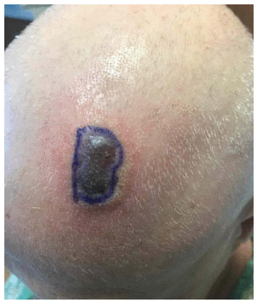
A purpuric bulla with surrounding erythema (ecthyma gangrenosum) on the scalp of a 55-year-old woman with cancer and neutropenic bacteremia caused by Pseudomonas aeruginosa. The base of the bulla is outlined by skin marker to define the extent of the lesion.
Ecthyma gangrenosum presents as skin lesions that begin as macules with surrounding erythema before rapidly progressing to bullae and necrotic, ulcerative eschars. Pathogenesis of ecthyma gangrenosum involves hematogenous seeding of the skin in the setting of bacteremia.1 Classically, the condition is diagnosed in patients who are immunocompromised with P. aeruginosa bacteremia. 2 Because of the strong association with Pseudomonas bacteremia, empiric intravenous antipseudomonal antibiotics are recommended when ecthyma gangrenosum is suspected.1 In patients with neutropenic fever, aspiration or biopsy of skin lesions is recommended. 1,3 In our patient's case, we deferred biopsy because her lesion resolved rapidly on empiric antibiotics. Although antibiotics should never be withheld when ecthyma gangrenosum is suspected, the differential diagnosis includes noninfectious lesions such as warfarin-induced skin necrosis, calciphylaxis and vasculitis.2
Footnotes
Competing interests: None declared.
This article has been peer reviewed.
The authors have obtained patient consent.
This is an Open Access article distributed in accordance with the terms of the Creative Commons Attribution (CC BY-NC-ND 4.0) licence, which permits use, distribution and reproduction in any medium, provided that the original publication is properly cited, the use is noncommercial (i.e., research or educational use), and no modifications or adaptations are made. See: https://creativecommons.org/licenses/by-nc-nd/4.0/

Comments
Post a Comment